Patient Guide
Being told you need surgery for a brain tumor is an emotionally overwhelming moment for any patient and their loved ones. To help answer your questions and face this process with greater peace of mind, this guide explains each stage of brain tumor surgery in detail, from the operating room to your recovery.
What is a craniotomy? It is the surgery used to remove most brain tumors. A section of the skull bone is temporarily removed to reach the tumor and, at the end, the bone is put back in place.
Does the surgery hurt? No. It is done under general anesthesia or, in selected cases, with the patient awake and the scalp numbed. The brain itself has no pain receptors.
How Long Does Recovery Take? As a general reference, the hospital stay is about 3 days, and most people return to their daily activities 2 to 4 weeks later.
Medically reviewed by Dr. José Raúl García Zamarrón, brain and spine neurosurgeon
In this guide
- What is a craniotomy?
- When is brain tumor surgery recommended?
- What are the types of craniotomy?
- How does the neurosurgeon choose the approach and how much tumor to remove?
- What technology is used during surgery?
- How do you prepare for surgery?
- How is the surgery performed, step by step?
- Why are some surgeries done with the patient awake?
- What happens during the hospital stay?
- What is recovery like at home?
- What are the risks of a craniotomy?
- What results can you expect?
- Key takeaways
- Frequently Asked Questions
What is a craniotomy?
A craniotomy is a surgery in which a window is opened in the skull bone to reach the brain. It is the procedure used to remove most brain tumors.
The name describes what is done: “cranio” (skull) and “-tomy” (cut). The piece of bone that is removed is called a bone flap, and it is kept during surgery so it can be put back at the end.
Besides tumors, a craniotomy is used to treat brain aneurysms, blood vessel malformations, hematomas, and other lesions inside the skull.
Craniotomy vs. craniectomy: not the same thing
In a craniotomy, the bone is put back at the end of surgery. In a craniectomy, the bone is not replaced right away, usually because the brain is swollen and needs room, as happens in some severe head injuries.
For planned brain tumor surgery, a craniotomy is the standard approach.
When is brain tumor surgery recommended?
Brain tumor surgery is recommended when the tumor causes symptoms, grows, presses on important structures, or its type must be confirmed to plan treatment. Not every tumor needs surgery: some can be monitored with regular MRI scans or treated with radiation therap
Surgery serves several goals at once:
- Remove the tumor: take out as much as possible without affecting brain function.
- Relieve pressure: reduce the pressure inside the skull, which can improve symptoms such as headache or drowsiness.
- Confirm the diagnosis: obtain tissue so the neuropathologist, a specialist in analyzing nervous system tissue, can determine the tumor’s type and grade.
- Guide further treatment: the results show whether radiation therapy, chemotherapy, or only follow-up is needed.
To learn about symptoms, tumor types, and other treatment options, read our guide on brain tumors: symptoms, types, diagnosis, and treatment.
When a craniotomy is not needed
Some tumors are treated through other surgical approaches.
Minimal invasiveness
Endoscopic endonasal surgery
Pituitary adenomas and certain skull base tumors can be removed with endoscopic endonasal surgery, a minimally invasive technique: the surgeon reaches the tumor through the nose with an endoscope, without opening the skull and with no visible incisions.
Diagnosis
Stereotactic biopsy
When a tumor is very deep and only its type needs to be confirmed, a stereotactic biopsy may be performed: a small sample is taken through a tiny opening, guided by three-dimensional coordinates.
To find out which approach is best for your case, you can request an evaluation with Dr. García Zamarrón, a neurosurgeon in Ciudad Juárez trained in Mexico, Spain, and Brazil.
Request ConsultationWhat are the types of craniotomy?
The type of craniotomy is chosen based on the tumor’s location. The goal is to reach it by the most direct route while disturbing as little healthy tissue as possible.
Modern neurosurgery favors openings that are only as large as needed. With the microscope and neuronavigation, in many cases it is possible to work through smaller craniotomies than in the past.
| Approach | Where the incision is made | Areas it is used for |
|---|---|---|
| Frontotemporal (pterional) | Behind the hairline, on one side of the head | Frontal and temporal lobes, anterior skull base |
| Supraorbital | In the eyebrow, through a small incision | Some lesions of the anterior skull base |
| Convexity | Over the tumor site, within the hairline | Tumors on the surface of the brain, such as many meningiomas |
| Retrosigmoid | Behind the ear | Tumors of the hearing nerve and the cerebellopontine angle |
| Suboccipital | At the back of the neck | Tumors of the cerebellum and the back of the skull |
How does the neurosurgeon choose the approach and how much tumor to remove?
The neurosurgeon chooses the approach and extent of surgery by seeking the best balance between removing as much tumor as possible and preserving brain function. In neurosurgery, this principle is known as maximal safe resection.
Criteria that define the approach
- Location and depth: determine the shortest route to the tumor and the patient’s position in the operating room.
- Relationship to eloquent areas: when the tumor is close to the areas for speech, movement, or vision, the approach is planned to avoid crossing them, and awake surgery is considered.
- Relationship to blood vessels and nerves: nearby arteries, veins, and cranial nerves determine the working angle and dissection technique.
- Suspected tumor type: a well-defined tumor, such as a meningioma, allows a different strategy from a tumor that infiltrates tissue, such as a glioma.
- Patient’s health: age, other medical conditions, and neurological status influence the extent and length of surgery.
What total or partial removal means
The extent of surgery is described by how much of the tumor is removed, and it is confirmed with a follow-up MRI:
| Extent | What it means | When it is chosen |
|---|---|---|
| Gross total resection | No visible tumor on the follow-up MRI | When the tumor can be safely separated from healthy tissue |
| Subtotal resection | Most of the tumor is removed, leaving a small remnant | When part of it is attached to structures that control important functions |
| Partial resection | Part of the tumor is removed to relieve pressure and obtain tissue | Large tumors or tumors in high-risk areas |
| Biopsy | Only a sample is taken for diagnosis | Deep tumors, or when the main treatment will be radiation therapy or chemotherapy |
How tumor type makes a difference
In gliomas, medical evidence links a greater extent of resection with better disease control, as long as neurological function is not compromised. That is why tools that help identify the tumor’s borders are used.
In meningiomas, the goal includes also removing the portion of membrane where the tumor began, when it is safe to do so, because this lowers the chance that it will come back. Neurosurgeons describe this relationship with the Simpson grading scale.
For metastases, surgery is usually recommended when there is a single or large lesion that can be reached, and it is followed by radiation therapy to the surgical area.
When it is safer to leave a remnant
If part of the tumor is attached to an area that controls speech, movement, or vital functions, it may be better to leave a small remnant and treat it later with radiation therapy or monitor it. Preserving the patient’s quality of life is a central part of surgical judgment.
What technology is used during surgery?
Brain tumor surgery today relies on technologies that act as layers of safety: they help plan the route, see the tumor’s borders, and protect brain function. Not all of them are used in every surgery; the neurosurgeon chooses the most appropriate ones based on the tumor’s type and location.
1 ·Planning
Artificial Intelligence
Creates three-dimensional maps that identify critical areas to avoid.
Neuronavigation
Shows the position of instruments relative to the tumor in real time, like a GPS for the brain.
Tractography
An MRI technique that shows the path of nerve fibers for movement and speech.
2 · Removal
Surgical microscope
Magnifies and lights the surgical field to tell the tumor apart from vessels and nerves.
Fluorescence-guided surgery
A substance makes the tumor’s borders visible under a special light.
Ultrasonic aspirator
Breaks up and suctions the tumor gradually and in a controlled way.
3 · Protection and diagnosis
Neuromonitoring and brain mapping
Tracks movement and sensory pathways and locates functional areas.
Intraoperative pathology
Rapid analysis of a sample while the patient is still in the operating room
How do you prepare for surgery?
Preparation begins days before surgery, with tests confirming that the patient can safely undergo the operation and with detailed surgical planning.
In the days before surgery
- Pre-operative tests: blood work and, depending on age and medical history, a cardiology or other specialist evaluation.
- Planning MRI: an MRI with special slices that is loaded into the neuronavigation system to map the route to the tumor.
- Medication review: some medications, such as blood thinners and certain anti-inflammatories, must be stopped before surgery. Adjust them only as directed by your doctor.
- Preparatory medications: some patients are prescribed corticosteroids to reduce brain swelling or anti-seizure medication to prevent seizures.
- Informed consent: the neurosurgeon explains the surgical plan, its benefits and risks, and answers questions from the patient and family.
On the day of surgery
The patient arrives at the hospital fasting, at the scheduled time. An IV line is placed, and the anesthesiologist performs a final evaluation.
In most cases, only a narrow strip of hair is shaved at the incision site, and sometimes no shaving is needed at all.
Helpful questions for your pre-surgery visit
- What type of tumor is suspected, and how much of it can be removed?
- Which functions could be affected because of the tumor’s location?
- Will I have general anesthesia or awake surgery?
- How many days will I be in the hospital, and when can I return to my activities?
- What treatment might I need after surgery?
How is the surgery performed, step by step?
Learn the main stages of a craniotomy to remove a brain tumor.
Step 1
Anesthesia and positioning
General anesthesia, except in awake surgery. The head is secured with a holder that keeps it completely still.
Step 2
Neuronavigation registration
The system matches the MRI to the patient’s actual anatomy, and the exact incision site is marked.
Step 3
Incision and skull opening
An incision is made within the hairline. Small holes (burr holes) are drilled and the bone flap is removed.
STEP 4
Opening the dura
The outermost membrane covering the brain is opened to expose the tumor area.
STEP 5
Tumor removal
Under the microscope and with the technologies suited to the case, the tumor is separated from healthy tissue. A sample is sent to pathology.
STEP 6
Inspection and closure
Any bleeding is controlled, the dura is closed, the bone is secured with titanium plates, and the skin is closed in layers.
STEP 7
Waking up and first assessment
The patient wakes up and is checked to make sure they can answer questions and move their arms and legs.
Duration and recovery
Reference times that may vary from case to case.
3 to 5 hours
Typical length of surgery.
About 3 days
Typical hospital stay.
2 to 4 weeks
Typical return to daily activities.
Recovery depends on the tumor type, its location, and each patient’s condition.
Why are some surgeries done with the patient awake?
Awake craniotomy is performed when the tumor is close to the brain areas that control speech or movement, known as eloquent areas. Keeping the patient awake during tumor removal makes it possible to confirm in real time that these functions are preserved.
The patient does not feel pain. The scalp is numbed with local anesthetic, and the brain has no pain receptors. During the opening and closing, the patient is sedated.
During tumor removal, the team asks the patient to do simple tasks: talk, count, name pictures, or move a hand. If a task is affected, the neurosurgeon knows they are near an important area and adjusts the removal.
Not every patient is a candidate. The decision takes into account the tumor’s location, neurological status, and the patient’s ability to cooperate during surgery, and it is prepared together with the patient during pre-surgery visits.
What happens during the hospital stay?
As a general reference, the hospital stay after a craniotomy for a brain tumor is about 3 days. On the first night, the patient stays in a close-monitoring unit.
- Neurological monitoring: the team frequently checks alertness, strength, speech, and vision.
- Pain control: incision pain is usually well controlled with pain medication.
- Early mobility: patients sit up and walk early, usually from the next day, which supports recovery.
- Follow-up MRI: a scan is done in the first few days to evaluate how much of the tumor was removed.
Swelling or bruising around the eye on the side of surgery is common, especially after surgery on the front and side of the head. It goes away on its own within a few days.
What is recovery like at home?
Craniotomy recovery at home is gradual: as a general reference, most people return to daily activities 2 to 4 weeks after surgery. Fatigue is common in the first few weeks and improves little by little.
Care during the first few weeks
- Activity: walking every day supports recovery; strenuous exercise and lifting are resumed once your neurosurgeon clears you.
- Incision: keep it clean and dry; stitches or staples are removed at your follow-up visit.
- Driving and medications: do not drive or stop corticosteroids or anti-seizure medication without your doctor’s approval.
Call 911, go to the emergency room, or call your neurosurgeon if you have:
- Fever, redness, or drainage from the incision
- A severe headache that gets worse instead of better
- A seizure
- New weakness, trouble speaking, or confusion
- Extreme drowsiness or persistent vomiting
What are the risks of a craniotomy?
Like any surgery, a craniotomy carries risks, which your neurosurgeon will review with you before the procedure based on the tumor’s location and your overall health.
General risks of any surgery
These include reactions to anesthesia, bleeding, wound infection, and blood clots in the leg veins, which are prevented with early mobility and other measures.
Risks specific to brain surgery
| Risk | What it involves |
|---|---|
| Brain swelling (edema) | May temporarily worsen some symptoms and is treated with medication. |
| Bleeding inside the skull | Occasionally requires another operation. |
| Seizures | May occur after surgery and are usually controlled with medication. |
| Cerebrospinal fluid leak | Leakage of the fluid that surrounds the brain through the incision |
| Deep infection | Such as meningitis, an infection of the membranes covering the brain. |
| Neurological deficit | Weakness or problems with speech, vision, or memory, which may be temporary or permanent. |
Planning with neuronavigation and neuromonitoring during surgery are intended to reduce these risks.
What results can you expect?
Results vary from case to case: they depend mainly on the tumor type, its location, and how much could be safely removed. For many benign tumors, complete removal provides long-term control; for malignant tumors, surgery is the first step of combined treatment.
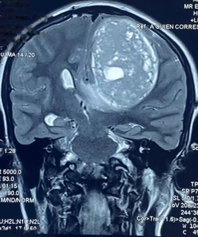 Before · coronal view
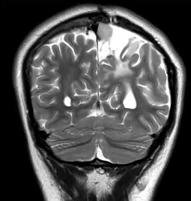 After · coronal view
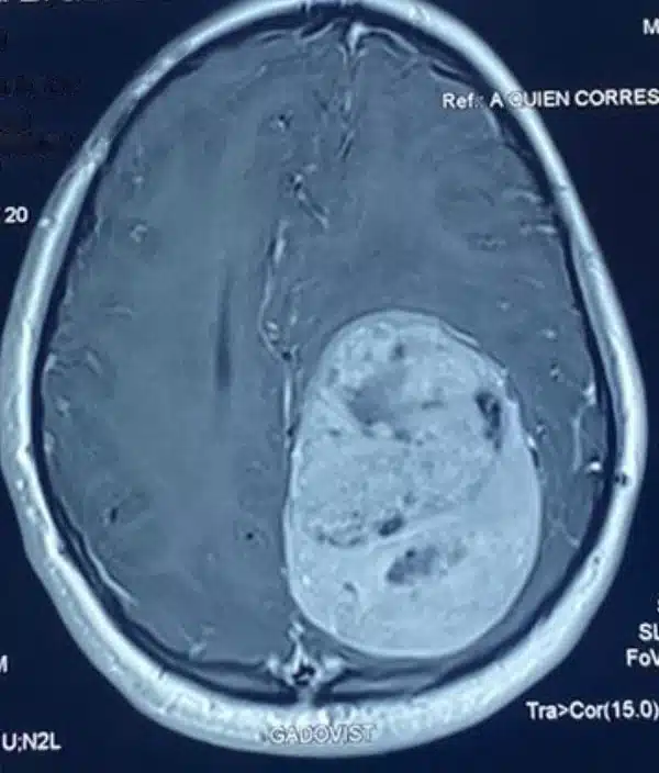 Before · axial view
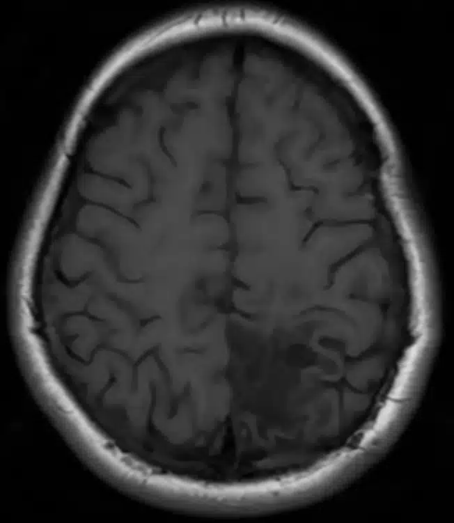 After · axial view
Some symptoms, such as headache caused by pressure, may improve within the first few days. Others, such as weakness or speech problems, improve gradually over weeks or months.
The pathology diagnosis and follow-up MRI scans help define the next steps in treatment and detect any change early.
Key takeaways
- A craniotomy is the surgery used to remove most brain tumors; the bone is put back at the end.
- The type of craniotomy is chosen based on the tumor’s location, seeking the most direct route with minimal impact on healthy tissue.
- Surgery aims for maximal safe resection: removing as much tumor as possible while preserving brain function.
- Neuronavigation, fluorescence, and neuromonitoring help remove the tumor while protecting brain function.
- When the tumor is near the areas for speech or movement, surgery may be performed with the patient awake.
- As a general reference, the hospital stay is about 3 days, and most people return to their activities 2 to 4 weeks later.
Frequently asked questions about craniotomy
Will my whole head be shaved?
No. In most cases, only a narrow strip of hair is shaved at the incision site, and sometimes no shaving is needed. As your hair grows back, the scar is usually hidden.
Is there a lot of pain after surgery?
Pain after a craniotomy is centered on the incision and the muscle in the surgical area, and it is usually controlled with pain medication. It eases steadily over the first few days.
Does the skull bone go back in place?
Yes. In a craniotomy, the piece of bone is put back at the end of surgery and secured with small titanium plates and screws. Over time, the bone heals. Titanium is safe for future MRI scans.
When can I go back to work?
As a general reference, most people return to daily activities 2 to 4 weeks after surgery. Jobs involving physical effort or operating machinery may require more time, and the date is set at your follow-up visit.
When will I know if the tumor was benign or malignant?
The final diagnosis comes from the pathology report, which is usually ready within days or weeks after surgery. That result determines whether any additional treatment is needed.
Is it possible that not all of the tumor is removed?
Yes. When part of the tumor is in contact with structures that control important functions, it may be safer to leave a small remnant and treat it later with radiation therapy or monitoring. The priority is to preserve the patient’s function.
Can I get a second opinion before surgery?
Yes. Getting a second opinion is your right and a common step before brain surgery. For your visit, bring your imaging studies in digital format and the radiology reports.
Consultation & Evaluation
Schedule Your Evaluation
If you have been told you need brain tumor surgery, a specialist evaluation gives you something concrete: understanding the surgical plan, the alternatives, and what to expect at each stage of recovery. Dr. García Zamarrón sees patients at Hospital Ángeles in Ciudad Juárez, just across the border from El Paso, Texas.
Schedule Your Consultation Message Us on WhatsAppDr. José Raúl García Zamarrón —Neurosurgeon · Brain & Spine
Av. Campos Elíseos 9371, Office 795, Col. Campos Elíseos, C.P. 32420, Ciudad Juárez, Chihuahua
Phone 656 227 1963 WhatsApp 656 657 7818
Monday to Friday, 9:00 AM–5:00 PM · Saturdays, 9:00 AM–12:00 PM
Keep Reading
Medical Review
Dr. José Raúl García Zamarrón
Neurosurgeon · Brain & Spine · Ciudad Juárez, Chihuahua
Mexican medical license: 6241285
Specialty license: 10138902
Certified by the Mexican Council of Neurological Surgery
Member of AO Spine and EANS
Learn about the specialist’s background →
Last updated: October 2026.
Medical disclaimer
This content is for informational and educational purposes only. It does not replace a medical consultation, diagnosis, or personalized treatment. If you have neurological symptoms, see a specialist. If they are sudden or severe, seek emergency medical care.